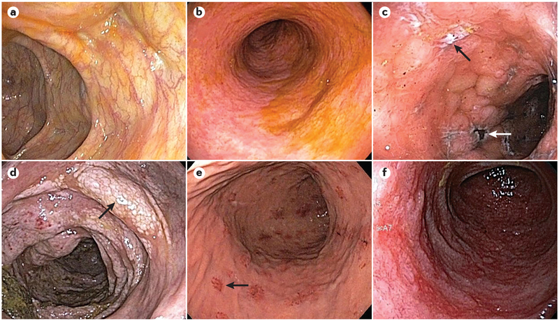
Figure 1 |. Endoscopic findings in acute GVHD of the gastrointestinal tract.
Endoscopic images depict various endoscopic findings in acute graft-versus-host disease (GVHD) of the gastrointestinal tract. a | Colonic mucosa with a normal appearance following haematopoietic stem cell transplantation (HSCT) in a patient with diarrhoea. Non-targeted biopsies revealed mild acute GVHD. b | Sigmoid colonic mucosa with mucosal oedema, loss of normal haustra and complete loss of vascularity following HSCT in a patient with diarrhoea. Biopsies revealed mild acute GVHD. c | Colonic mucosa with oedema, exudates, deep ulcers (black arrow) and areas of necrotic tissue (white arrow) in a patient following HSCT with biopsies confirming severe acute GVHD. d | Colonic mucosa with oedema, friability, loss of vascularity and white plaques (arrow) in a patient following HSCT. Biopsies revealed moderate acute GVHD and pneumatosis intestinalis. e | Upper endoscopy in a patient with nausea and epigastric pain following HSCT, showing patchy, raised erythematous lesions (arrow) as well as localized superficial mucosal erosions. Biopsies of the lesions revealed mild acute GVHD. f | Duodenal mucosa in a patient with severe epigastric pain following HSCT, revealing loss of normal plicae circulares, the presence of friable and oedematous mucosa and a complete loss of normal vascularity. Biopsies revealed severe acute GVHD.

