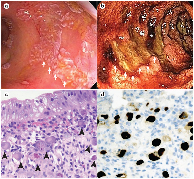
Figure 4 |. Gastrointestinal cytomegalovirus infection following HSCT.
Endoscopic and histopathological images from patients with acute graft-versus-host disease (GVHD) of the gastrointestinal tract, depicting cytomegalovirus infection in the post-haematopoietic stem cell transplantation (HSCT) setting. a | Colonoscopy in a patient following HSCT with established severe acute GVHD who is unresponsive to corticosteroids. Numerous raised white plaques (white arrows) are present throughout the colon. Biopsy samples of the lesions revealed extensive cytomegalovirus-infected cells and positive immunostaining for cytomegalovirus proteins. b | Colonoscopy in a patient following HSCT with severe acute GVHD and profuse haematochezia. Large, deep ulcers in the transverse colon (white arrows, which trace the rim of an ulcer), areas of active bleeding (asterisk) and diffusely oedematous colonic mucosa are seen. Biopsies of the mucosa revealed acute GVHD, whereas biopsies of the ulcer base stained positive for cytomegalovirus proteins by use of immunohistochemistry. c | Haematoxylin and eosin (H&E)-stained slide showing an area of a colon with crypt loss and multiple cytomegalovirus-infected endothelial cells (arrowheads, ×200). The cytomegalovirus-infected cells have large, smudgy, eccentric nuclei with prominent intranuclear and intracytoplasmic inclusions. d | Immunohistochemical staining of cytomegalovirus proteins (typically cytomegalovirus tegument component pp65) highlights cytomegalovirus-infected endothelial cells (dark brown immunostaining, ×200).

