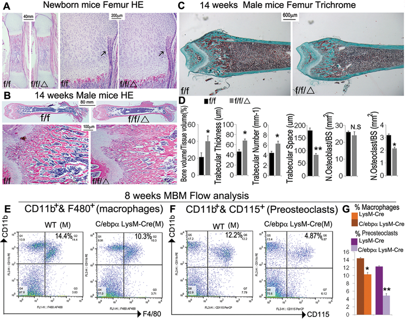
Figure 2. C/ebpαf/fLysM-Cre mice showed significantly impaired OC function, with mild changes in macrophage development and a large reduction in preosteoclast number.

(A) Representative images of H&E staining of (A) newborn and (B) 14-week-old C/ebpαf/fLysM-Cre (f/f/Δ) femurs with C/ebpαf/f (f/f) and LysM-cre as controls with high magnification of the epiphysis regions (n=10). (with 6 male and 6 female in each group). (C) Representative images of Goldner’s Trichrome analysis and (D) quantification data for male 14-week-old C/ebpαf/fLysM-Cre compared to C/ebpαf/f mouse femurs (n=16), which reconfirmed the severe osteopetrotic phenotypes in C/ebpαf/fLysM-Cre while osteoblast and OC numbers of mutants and wild-type remained almost unchanged. Analyses were repeated for female mouse samples with similar findings. (E, F) Flow cytometry for (E) immune cell subtyping of CD11b+ F4/80+ macrophages and (F) CD11b+CD115+ OCs from 8-week-old mouse bone marrow. (G) Quantification of E and F. Results show means ± SD. of triplicate independent samples. *p<0.05, **p<0.01. N.S., not significant.
