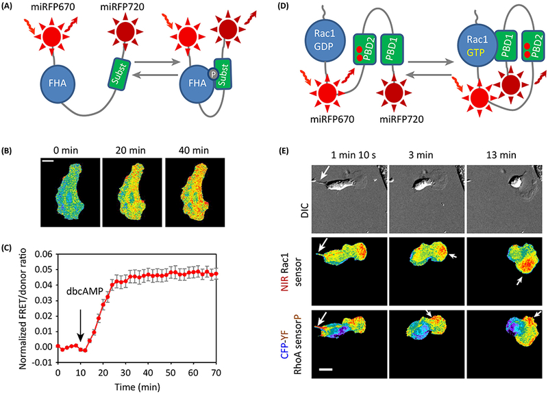
Figure 2. Single-chain NIR biosensors for different targets in signaling cascades based on miRFP670-miRFP720 FRET pair.

(A) Schematics of the NIR FRET biosensors for AKAR PKA and JNKAR JNK kinases. FHA is the phosphopeptide binding domain that recognizes phosphorylated peptide marked as “subst“. (B,C) NIR AKAR PKA kinase biosensor was validated in live HeLa cell stimulated with 1 mM dibutyryl cAMP (dbcAMP). Time-lapse images (B) and the corresponding plot (C) are shown. (D) Schematics of the FRET Racl biosensor. PBD1 is a p21-binding domain 1, PBD2 is a mutant p21-binding domain. Racl is a full-length Racl post-translationally isoprenylated for membrane localization. (E) Combination of NIR and cyan-yellow FRET biosensors for simultaneous imaging of Racl and RhoA activities in a MEF/3T3 cell. DIC, differential interference contrast. Racl is predominantly localized at the leading edge, whereas RhoA activity is mostly localized at the retracting tail, the side edges and at the back of the leading-edge protrusions (see arrows). (B,E) FRET/donor ratio is shown in pseudocolor. Scale bar, 20 μm. (C) Mean ±s.e.m. (n=3) of the FRET/donor ratio for the whole cell plotted vs time. B,C,E adapted with permission from [19]
