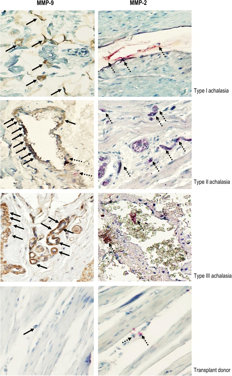
Fig. 3. Immunohistochemical analysis of MMP-2 and MMP-9.
Representative immunostaining of MMP-9 (left panel) and MMP-2 (right panel) in tissue biopsies from achalasia patients. Solid arrows depict MMP-9 expression (in brown). Dotted arrows show MMP-2 expression (in red). Original magnification was X320. Below the achalasia tissue samples, a normal biopsy from the esophagus of a transplant donor is included. The immunoreactivity for MMP-9 was with the monoclonal antibody REGA-2D9, whereas the immunoreactivity for MMP-2 was with a polyclonal antibody

