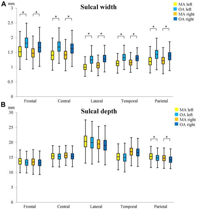
Figure 3.

Differences in sulcal width (A) and depth (B) between middle-age (MA) and old-age (OA). The median, 1st and 3rd quartile range of sulcal widths and depths in MA and OA are shown. Whiskers show minimum and maximum value. *Indicates significant (p < 0.01) age group differences in sulcal width/depth. Superior frontal sulcus (frontal); central sulcus (central), lateral sulcus (lateral); superior temporal sulcus (temporal); intra-parietal sulcus (parietal).
