
Figure 3.
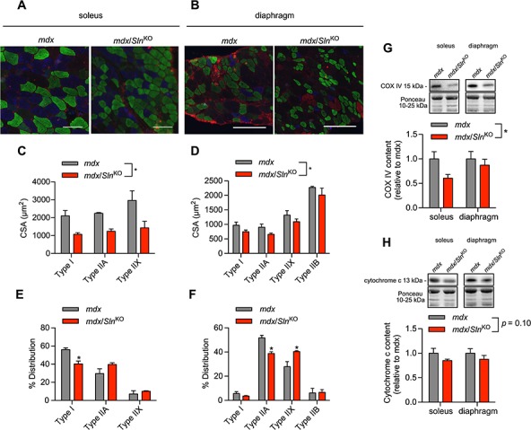
Immunofluorescent fibre typing (A and B) revealed a significant reduction in myofibre CSA (C and D) and a slow-to-fast fibre-type shift (E and F) in response to Sln deletion in the mdx mice (n = 3−4 per group). Western blotting demonstrated a significant reduction in COX IV (G) and a trending reduction in cytochrome c (H) in muscles from mdx/SlnKO mice compared with mdx. For (C), (D), (G) and (H), *denotes a significant main effect when using a two-way ANOVA, P ≤ 0.05.for (E) and (F), *denotes significance using a Student’s t-test comparing mdx versus mdx/SlnKO muscles, P ≤ 0.05.