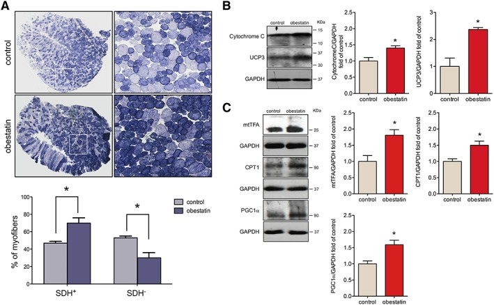
Figure 2.

Obestatin treatment increases oxidative fibre density. (A) Upper panel, representative succinate dehydrogenase (SDH) staining from tibialis anterior (TA) from control muscles and obestatin‐treated muscles at 30 days. Bottom panel, quantitation of SDH+ and SDH− muscle fibres from TA muscles. Data were expressed as mean ± SEM (* P < 0.05 vs. control values). (B) Immunoblot analysis of Cytochrome C and uncoupling protein 3 (UCP3) in TA muscles after intramuscular injection of obestatin or control (phosphate‐buffered saline) at 30 days. (C) Immunoblot analysis of mitochondrial transcription factor A (mtTFA), carnitine palmitoyltransferase‐1 (CPT1), and proliferator‐activated receptor‐gamma coactivator 1α (PGC1α) in TA muscles after intramuscular injection of obestatin or control (phosphate‐buffered saline) at 30 days. In (B) and (C), protein level was expressed as fold of control TA muscles, and immunoblots are representative of the mean value. Data were expressed as mean ± SEM obtained from intensity scans (* P < 0.05 vs. control values).
