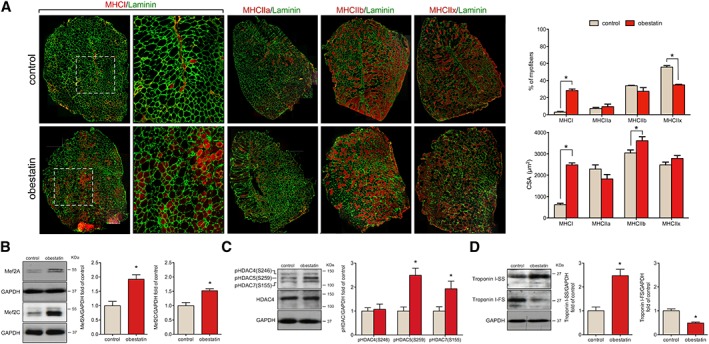
Figure 3.

Obestatin stimulates fast to slow twitch fibre type shifting in mdx mice. (A) Left panel, representative images of control‐treated and obestatin‐treated tibialis anterior (TA) muscles showing myosin heavy chains (MHC) expression. Mice muscle serial cross section incubated with a primary antibody against MHCI, MHCIIa, MHCIIb, or MHCIIx, followed by incubation with appropriate fluorescent‐conjugated secondary antibody. Right panel, quantitation of fibre types (upper panel) and cross‐sectional area (CSA) (bottom panel). Data are shown as mean ± SEM of five animals per group (* P < 0.05 vs. control values). (B) Immunoblot analysis of Mef2A and Mef2C in TA muscles after intramuscular injection of obestatin or vehicle (phosphate‐buffered saline, control) at 30 days. (C) Immunoblot analysis of pHDAC4(S246), pHDAC5(S259), pHDAC7(S155), and HDAC4 in TA muscles after intramuscular injection of obestatin or control (phosphate‐buffered saline) at 30 days. (D) Expression of the slow‐fibre‐specific troponin I‐SS and fast‐fibre‐specific troponin I‐FS in TA muscles after intramuscular injection of obestatin or vehicle at 30 days. In (B) to (D), protein level was expressed as fold of control TA muscles, and immunoblots are representative of the mean value. Data were expressed as mean ± SEM (n = 5 per group) obtained from intensity scans (* P < 0.05 vs. control values). HDAC, histone deacetylases; Mef2, myocyte enhancer factor‐2.
