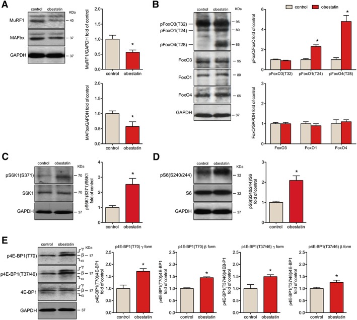
Figure 4.

Obestatin treatment protects from atrophy with induction of protein synthesis signalling in mdx mice. (A) Immunoblot analysis of MuRF1 and MAFbx in tibialis anterior (TA) muscles after intramuscular injection of obestatin or control. (B) Immunoblot analysis of the phosphorylation partner of FoxO3, FoxO1, and FoxO4 in TA muscles after intramuscular injection of obestatin or vehicle (phosphate‐buffered saline, control) at 30 days. (C) Analysis of pS6K1(S6371) and S6K1 in TA muscles after intramuscular injection of obestatin or vehicle at 30 days. (D) Immunoblot analysis of pS6(S240/244) and S6 in TA muscles after intramuscular injection of obestatin or vehicle. (E) Analysis of p4E‐BP1(T70), p4E‐BP1(T37/46), and 4E‐BP1 in TA muscles after intramuscular injection of obestatin or vehicle (phosphate‐buffered saline, control) by immunoblot. In (A) to (E), protein level was expressed as fold of control TA muscles. Immunoblots are representative of the mean value (n = 5 per group). Data were expressed as mean ± SEM obtained from intensity scans (* P < 0.05 vs. control values).
