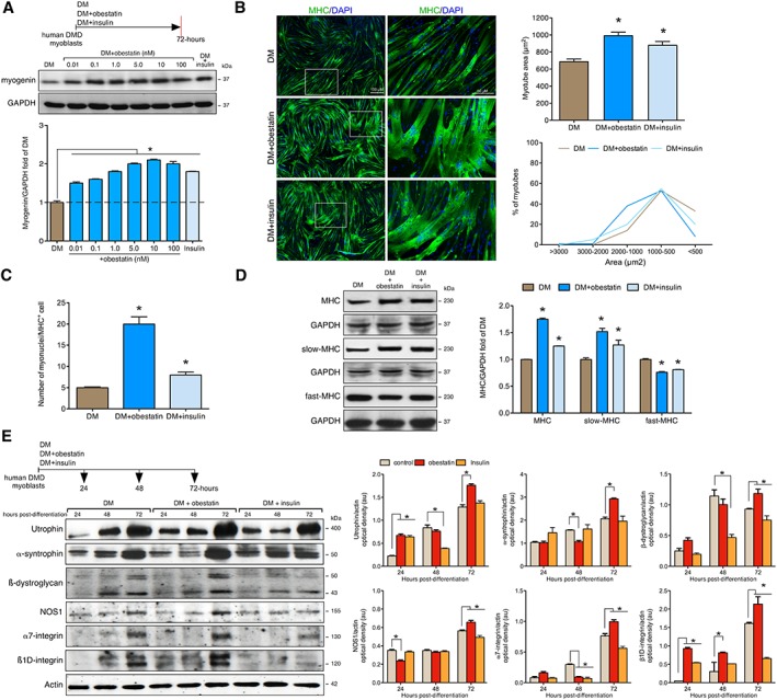
Figure 7.

Obestatin signalling regulates differentiation of human Duchenne muscular dystrophy (DMD) cells. (A) Dose–response effect of obestatin (0.01–100 nM) or insulin (1.72 μM) on differentiating human DMD cells. Levels of myogenin were represented as a fold of respective expression in differentiation medium (DM) (72 h post‐differentiation). (B) Left panel, immunofluorescence detection of myosin heavy chains (MHC) and 4′,6‐diamidino‐2‐phenylindole (DAPI) in human DMD myotube cells under DM (control), DM + obestatin (10 nM), or DMD + insulin (1.72 μM) at the 72 h point after stimulation. Right panel, the differentiation grade was evaluated based on the myotube area and distribution. Data were expressed as mean ± SEM (* P < 0.05 vs. control values). (C) Quantification of the number of myonuclei in MHC+ cells in human DMD myotube cells under DM (control), DM + obestatin (10 nM), or DMD + insulin (1.72 μM) at the 72 h point after stimulation. Data were expressed as mean ± SEM (* P < 0.05 vs. control values). (D) Immunoblot analysis of MHC, slow‐MHC and fast‐MHC, in human DMD myotubes at the 72 h point under DM (control), DM + obestatin (10 nM), or DMD + insulin (1.72 μM). (E) Effect of obestatin (10 nM) on utrophin, α‐syntrophin, ß‐dystroglycan, nitric oxide synthase (NOS1), α7‐integrin, and ß1D‐integrin expression on differentiating human DMD cells. In (A), (D), and (E), protein level was expressed as optical density arbitrary units obtained from intensity scans. Immunoblots are representative of the mean value. Data were expressed as mean ± SEM (* P < 0.05 vs. control values).
