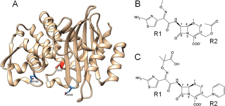
Figure 1.
Structures of CTX-M-14 β-lactamase and oxyimino-cephalosporins. A, ribbon diagram of CTX-M-14 β-lactamase. The Asn-106 and Asp-240 residues are highlighted in blue. The catalytic Ser-70 residue is highlighted in red. B, schematic illustration of cefotaxime. C, schematic illustration of ceftazidime.

