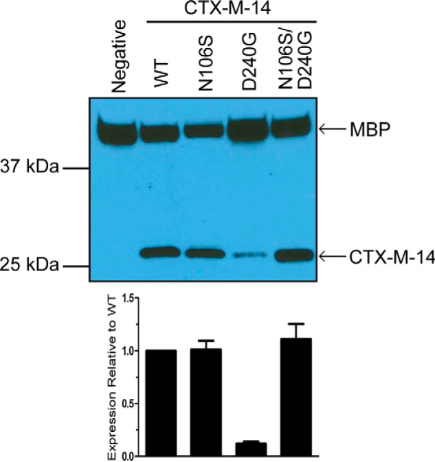
Figure 3.

Steady-state protein levels of WT CTX-M-14 and the mutant β-lactamases. Western blot analysis with an anti-CTX-M-14 polyclonal antibody shows protein expression levels in the periplasmic fraction of E. coli encoding WT CTX-M-14 and the N106S, D240G, and N106S/D240G mutant enzymes. Polyclonal antibody to the periplasmic protein MBP was used as a loading control. The signal was visualized by chemiluminescence and quantified by densitometry. The signal for CTX-M-14 β-lactamase was normalized to that for MBP in the same sample. The lower panel shows protein levels relative to the WT enzyme. The error bars represent the standard deviation of protein levels based on three experiments.
