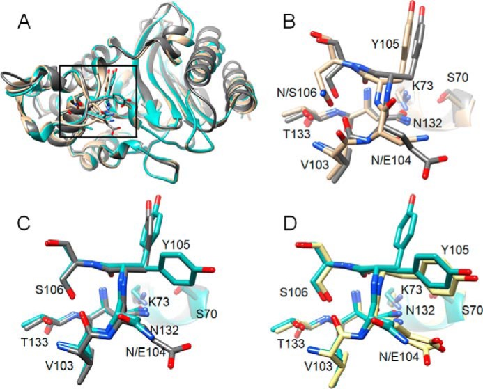
Figure 8.

Structural alignments of CTX-M-14, CTX-M-14 N106S, and TEM-1 β-lactamases. A, ribbon diagram of alignment of structures of WT CTX-M-14 (PDB code 1YLT) (9) (tan), N106S (blue-green), and TEM-1 (PDB code 1XPB) (dark gray). Side chains for residues 103–106, 132–133, and 70, 73 are shown as sticks. Box indicates the region of the structure shown in B–D. B, alignment of residues 103–106, 132–133, and 70, 73 for WT CTX-M-14 (tan) and TEM-1 (dark gray). C, alignment of residues 103–106, 132–133, and 70, 73 for CTX-M-14 N106S (blue-green) and TEM-1 (dark gray). D, alignment of residues 103–106, 132–133, and 70, 73 for CTX-M-14 N106S (blue-green) and TEM-1 V216AcrF mutant (PDB code 4ZJ1) (39) (yellow).
