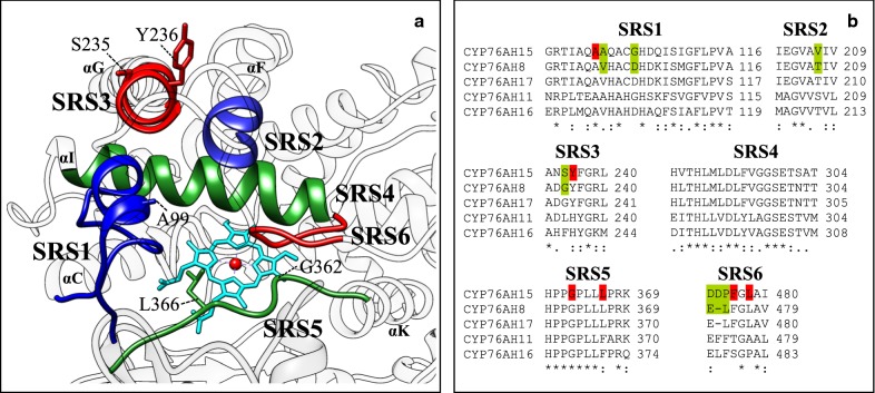
Fig. 2.
The SRS regions of selected CYP76AHs from Coleus forskohlii. a Homology model of CYP76AH15 with visualized SRS regions and selected helix-letters. SRS1, blue (left); SRS2, blue (right); SRS3, red (top); SRS5, green (bottom); and SRS6, red (right) were chosen as targeted areas for mutagenesis. The position of SRS4 on αI is shown in green. b Sequence alignments of the defined SRS regions of selected CYP76AH enzymes. Sites for mutagenesis are highlighted: Red; previously identified sites in other CYPs shown to confer changes in CYP properties (e.g. the SRS5 residues) or residues chosen in this work due to their structural positions. Green; sites of reciprocal mutagenesis of CYP76AH15 in comparison to CYP76AH8

