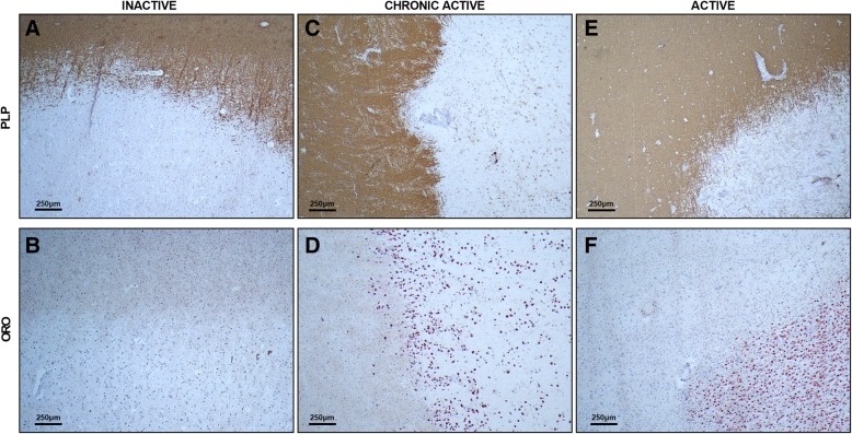
Fig. 1.
Histopathology of inactive, chronic active, and active multiple sclerosis lesions. Inactive, chronic active, and active multiple sclerosis (MS) lesions were stained for intracellular lipid droplets (oil red o; ORO) and myelin (proteolipid protein; PLP). a and b, c and d, e and f are taken from the same lesion. Foamy phagocytes (ORO+ cells) are apparent in demyelinating chronic active and active MS lesions, but not in inactive lesions

