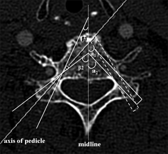
Figure 1.

Axial computed tomography images of the third cervical pedicle (C3) in a 74-year-old woman. The image shows the schematic diagram of pedicle transverse angles of C3 measured with the single line method and with the double-line method (analog nailing, 4.0 mm in diameter), respectively. In the diagram, a1 (or β1) and a2 (or β2) mean the minimum and maximum angles, respectively. Pedicle transverse angle values measured with single-line method shows a greater error range, and the accuracy is poor which does not meet the requirements of clinical practice in pedicle screw placement. While pedicle transverse angle values obtained with the parallel double-line measurement approach (i.e., double-line mimics the pathway of screw entry) could be more accurate and reliable, and the error range is significantly reduced, so the technique is more feasible for clinical operation
