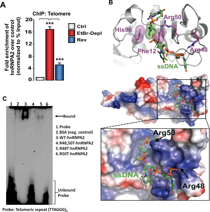
Fig 4. Identification of hnRNPA2 –telomere DNA binding domain.
(A) Association of hnRNPA2 at telomeric DNA assessed by ChIP assay in control, MtDNA-depleted, and MtDNA-depleted/hnRNPA2sh C2C12 cells using hnRNPA2 antibody. (B) Model of hnRNPA2 in complex with single stranded telomere DNA. The hnRNPA2 model has a basic groove postulated to bind ssDNA (green), with a clear pocket for the adenosine ring and key interactions with Arg48 and Arg50. The electrostatic surface potential map was generated with Delphi [47] and is colored from -7 kTe-1 (blue) to +7 kTe-1 (red). (C) EMSA showing telomere binding of hnRNP proteins using purified hnRNPA2 WT as well as hnRNPA2 KAT mutant proteins.

