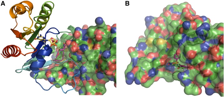
Figure 9.
ES4 Docking Simulations.
(A) Model of the crystal structure 1RE0 of yeast ARF1 (cartoon colored from the N terminus [blue] to the C terminus [red]) containing GDP (sticks, yellow) and a Mg ion (green sphere) with the GEA1-SEC7 domain (cartoon and transparent surface) in complex with BFA (sticks, purple) residing in a cavity at the ARF1-SEC7 interface. Shown is a position of ES4 (sticks, green) obtained by local docking in this cavity. ARF1’s switch-1 element loop (light-blue tube) occupies a hydrophobic groove of the SEC7 domain.
(B) Model of the crystal structure 4JWL of the human ARNO SEC7 domain (cartoon and transparent surface, similarly orientated as the SEC7 domain in [A]) in complex with N-(4-hydroxy-2,6-dimethylphenyl) benzenesulfonamide (sticks, purple). With blind dockings, ES4 (sticks, green) prefers to dock in the same depression where 4JWL’s ligand resides; this depression lies along SEC7’s hydrophobic groove that is recognized by an ARF switch-1 element loop.

