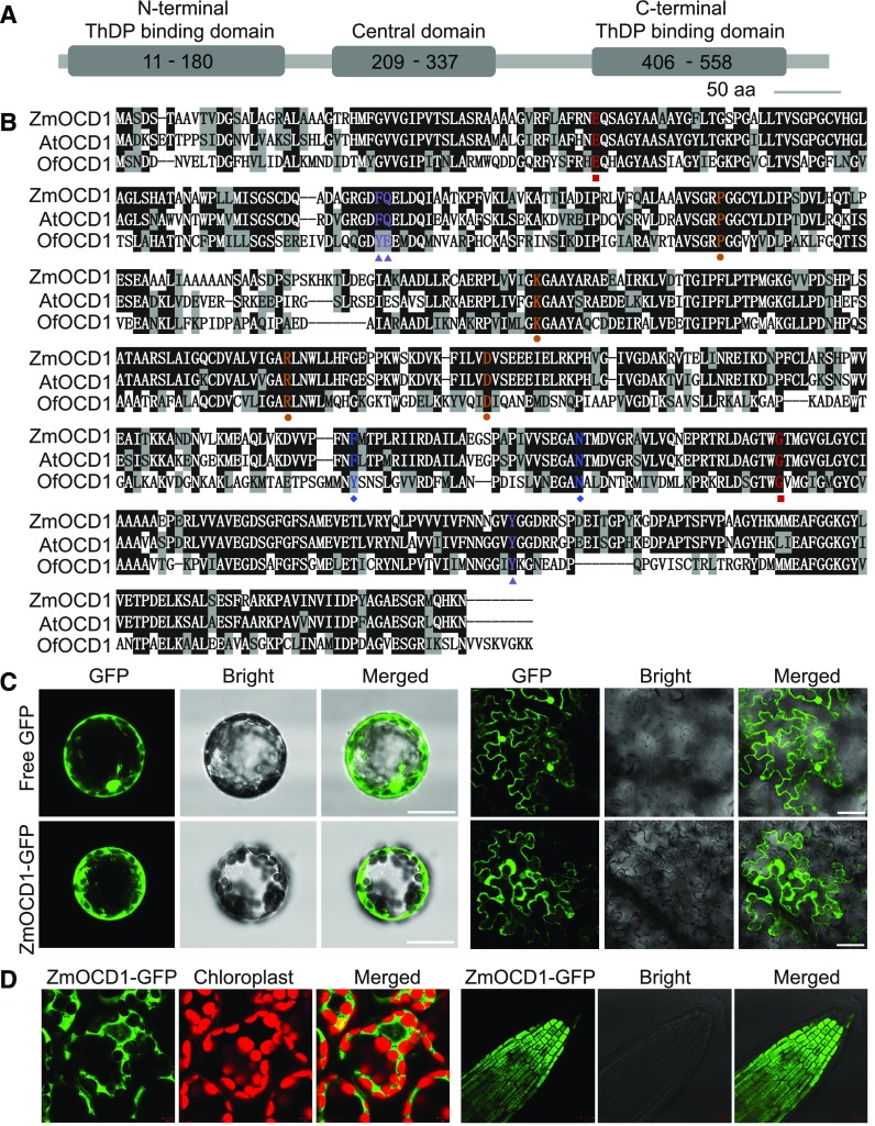
Figure 5.
Domain, Sequence, and Subcellular Localization of ZmOCD1.
(A) Schematic of the ZmOCD1 protein with conserved domains indicated. aa, amino acids.
(B) Alignment of the ZmOCD1 protein and its homologs from Arabidopsis and O. formigenes. The key amino acids for enzyme functions are indicated by different colored shapes. Red squares, ThDP binding sites; orange circles, ADP binding sites; blue diamonds, Mg binding sites; and purple triangles, active-site residues.
(C) Transient expression of the free 35S:GFP and 35S:ZmOCD1-GFP fusion (left panel) in Arabidopsis mesophyll protoplast cells and in tobacco leaf epidermal cells (right panel). Bars = 25 µm.
(D) Subcellular localization of ZmOCD1 in 35S:ZmOCD1-GFP transgenic plants. The GFP fluorescence in cotyledon (left panel) and root tip (right panel) cells from the T2 plants was observed.

