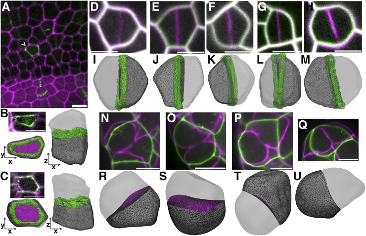
Figure 4.
Division Plane Prediction in Plant and Animal Cells.
(A) Micrograph of maize developing ligule. Cell walls are stained with PI (magenta) and division sites are marked by TAN1-YFP (green). Arrowhead indicates periclinal division.
(B) and (C) Micrographs of cells from the developing ligule expressing PIP2-CFP (magenta) for identification of cell outlines and TAN1-YFP (green) for division site location. The PPB and 3D reconstruction in both the XY (bottom left) and XZ plane (right) show the periclinal division plane.
(D) to (H) Time-lapse imaging of Arabidopsis guard cell division. The time point before the start of cytokinesis (green) overlaid with the completed division location (magenta).
(I) to (M) 3D reconstruction of cells in (D) to (H) with the corresponding predicted division plane by Surface Evolver. The final division site was the newly formed cell wall (green) for comparison to the predicted division.
(N) to (Q) Predicted division planes of cells in early gastrulation-stage C. elegans embryos. The cell shape prior to furrow ingression (green) was used for division predictions and overlaid with the dividing cell (magenta).
(R) to (U) 3D reconstruction of cells in (N) to (Q) along with the corresponding predicted division plane by Surface Evolver. Bars = 10 µm.

