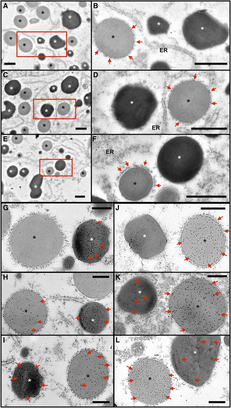
Figure 4.
Distribution of Glutelin and Prolamine Proteins in PB-I and PSV of P1MH and P3MH Endosperm Cells Compared with That of the Wild Type.
(A) to (F) Ultrastructure of PB-I and PSV in wild-type ([A] and [B]), P1MH ([C] and [D]), and P3MH ([E] and [F]) endosperm cells. (B), (D), and (F) are enlargements of the boxed regions in (A), (C), and (E). Bar = 1 μm. Red arrows denote rough ER surrounding PB-I. White asterisks indicate PSV, and black asterisks indicate PB-I.
(G) to (L) Immunolabeling of glutelin ([G] to [I]) and prolamine ([J] to [L]) proteins using monospecific antibodies and 15-nm gold particle-conjugated secondary antibodies. (G) and (J), wild type; (H) and (K), P1MH; (I) and (L), P3MH. Red arrows denote gold particle labeling. Note that in wild-type endosperm cells, glutelin and prolamine are dominantly detected in PSV and PB-I, respectively, although a few gold particles are evident due to slight background nonspecific labeling. Bar = 500 nm.

