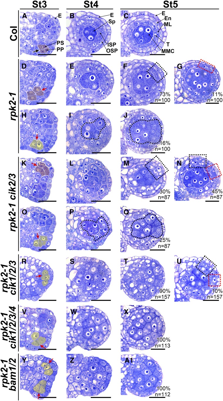
Figure 7.
CIKs Genetically Interact with RPK2 to Coordinate Cell Specification during Early Anther Development.
(A) to (C) Wild-type anthers at stage 3 (St3) (A), St4 (B), and St5 (C).
(D) to (J) Anthers of rpk2-1. At St3, some archesporial cells periclinally and symmetrically divide once to produce primary parietal cell- and primary sporogenous cell-like cells (shaded in red) (D). The primary parietal cell-like cells can divide once to produce two layers of secondary parietal cell-like cells at St4 (E). At St5, the MMC-like cells are enclosed by the tapetum-like cells and the endothecium-like cells (dotted black rectangle) (F) or by only one parietal cell layer (dotted red rectangle) (G). The other archesporial cells divide anticlinally (shaded in yellow), and no primary parietal cells can be produced (H), resulting in sporogenous cell-like cells (I) and MMC-like cells (J) enclosed only by the epidermis.
(K) to (Q) Anthers of rpk2-1 cik2/3 showing phenotypes similar to rpk2-1. In 75% of locules, one or two parietal cell layers can be produced at St5 (dotted red or black rectangle) ([M] and [N]). In 25% of locules, the MMC-like cells (dotted loops) are enclosed only by the epidermis (Q).
(R) to (U) Anthers of rpk2-1 cik1/2/3. Ninety percent of rpk2-1 cik1/2/3 locules produce MMC-like cells enclosed only by the epidermis (T). The other locules are enclosed by one or two parietal cell layers (dotted red or black rectangle) (U).
(V) to (X) rpk2-1 cik1/2/3/4 anthers showing phenotypes similar to bam1/2.
(Y) to (A1) rpk2-1 bam1/2 anthers showing phenotypes similar to bam1/2.
Thick black arrow indicates normal periclinal division of cells shaded in red (A). Red arrows indicate abnormal periclinal division of cells shaded in red ([D] and [K]) or anticlinal division of cells shaded in yellow ([H], [O], [R], [V], and [Y]). Sp-like cells ([I] and [P]) and MMC-like cells ([J] and [Q]) are indicated with dotted loops. Percentages of each phenotype are indicated. n indicates the total number of examined locules. E, epidermis; En, endothecium; ISP, inner secondary parietal cell; ML, middle layer; OSP, outer secondary parietal cell; PP, primary parietal cell; PS, primary sporogenous cell; Sp, sporogenous cell; T, tapetum. Bars = 20 µm.

