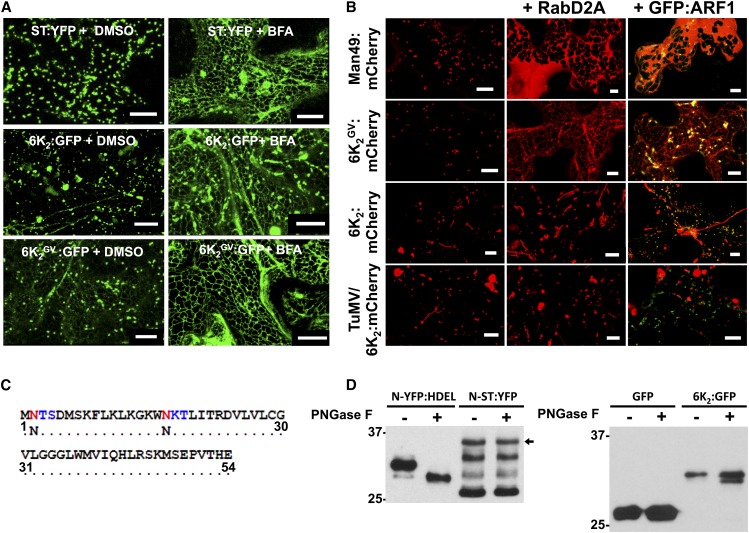
Figure 2.
Differential Sensitivity of 6K2 and 6K2GV to Inhibitors of ER-Golgi Trafficking.
(A) Confocal microscopy observation of N. benthamiana epidermal leaf cellsexpressing the trans-Golgi markers ST:YFP, 6K2:GFP, and 6K2GV:GFP were treated with DMSO (left panels) or with 20 μg/mL BFA (right panels) 24 h prior to confocal observation. Images are three-dimensional rendering stacks of 40 1-μm-thick slices that overlap by 0.5 μm.
(B) Confocal microscopy observation of N. benthamiana epidermal leaf cellexpressing the Golgi markers Man49:mCherry, 6K2GV:mCherry, and 6K2:mCherry ectopically or during TuMV infection alone (left panels) or in cells expressing Rab-D2A N123I (middle panels) or GFP:ARF1 NI (right panels). Images are three-dimensional renderings of 40 1-μm-thick slices that overlap by 0.5 μm.
(C) 6K2 N-glycosylation sites on the Asn residues in positions 2 and 17 (red) predicted by in silico analysis, forming N-X-S and N-X-T motifs (X-S/T in blue), respectively.
(D) Immunoblot performed with anti-GFP serum and protein extracts from N. benthamiana leaves expressing N-YFP:HDEL, N-ST:YFP, GFP, or 6K2:GFP that were treated with or without PNGase F. Black arrow indicates the position of N-ST:YFP. Two independent biological replicates were performed for (A) and (B), and three for (D). Bars = 20 μm in (A) and 10 μm in (B).

