Figure 8.
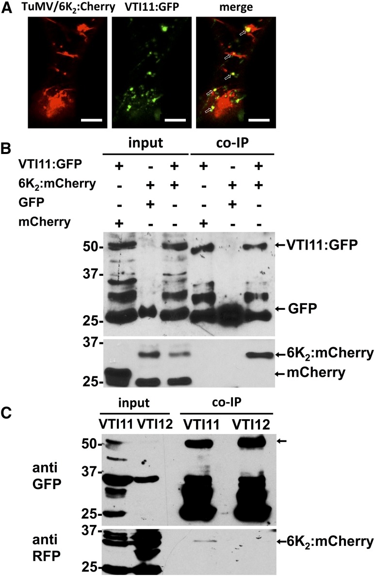
Interaction of the PVC SNARE VTI11 with 6K2.
(A) Confocal microscopy of N. benthamiana epidermal leaf cells expressing VTI11:GFP (middle panel) and 6K2:mCherry produced during TuMV infection (left panel). Colocalization of VTI11 with 6K2 is indicated by arrows in the merged image (right panel). Confocal images are single optical images of 1 μm thick.
(B) N. benthamiana leaves expressing combinations (upper table and indicated by arrows) of mCherry with VTI11:GFP, 6K2:mCherry with GFP, or 6K2:mCherry with VTI11:GFP were harvested 3 d after agroinfiltration. The cleared lysates (input) were used for co-IP and detection as described in Methods.
(C) N. benthamiana leaves expressing 6K2:mCherry with VTI11:GFP or VTI12:GFP were harvested 3 d after agroinfiltration. The cleared lysates were coimmunopurified as described in Methods.
Three independent biological replicates were performed for (A), four for (B), and two for (C). Bars = 10 μm in (A).

