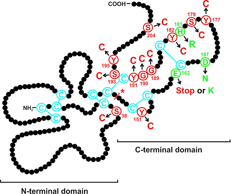
Figure 1:

Schematic illustration of the TIMP3 protein displaying mutations that cause autosomal dominant Sorsby fundus dystrophy. Each amino acid residue of the mature protein is shown by a black filled circle. The twelve cysteine residues predicted to form six disulphide bonds are indicated in blue. Mutations leading to an unpaired cysteine are shown in red, the three missense mutations leading to amino acid exchanges other than cysteines are depicted in green. An asterisk indicates the splice mutation. The figure has been modified from Figure 10 in Gliem et al., 2015, copyright holder: Association for Research in Vision and Ophthalmology
