Figure 2:

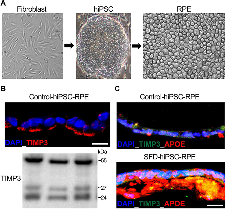
hiPSC-RPE as a tool to study SFD pathophysiology. A) Representative images showing patient-derived fibroblasts p.(Ser204Cys), hiPSCs and consequent differentiation of hiPSCs to obtain hiPSC-RPE monolayer. (B) Immunocytochemical and Western blotting analyses showing robust expression of TIMP3 in the basement membrane/ECM underlying control hiPSC-RPE cultures. Of note, the bottom panel shows the presence of TIMP3 bands consistent with unglycosylated, glycosylated and dimer forms of TIMP3 in ECM extracted from hiPSC-RPE cultures derived from three distinct control hiPSC lines. (C) Representative Immunoflurosecence labeling showing presence of APOE and TIMP3 positive drusen-like deposits underlying control vs. SFD hiPSC-RPE monolayer.
