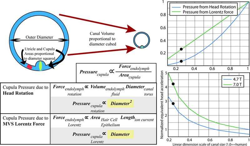
Figure 6.

Hypothesis for increased velocity of mouse eye movements in the magnetic field relative to prior reports in humans. Laboratory mice have much greater slow-phase nystagmus velocities than humans, despite a weaker magnetic field. This may be explained by differential scaling of the cupula pressures with labyrinth size due to a Lorentz force relative to a head rotation. For head rotations, the torque force due to the moment of inertia of the endolymph is related to the endolymph volume and the canal diameter. Therefore, cupula pressure due to head rotation is proportional to the diameter of the canal squared. For magnetic vestibular stimulation (MVS), the Lorentz force in the endolymph is related to the ionic current flowing into the area of hair cells, and to the length over which the current flows. Therefore, cupula pressure due to MVS is directly proportional to canal diameter. The right panels are graphical representations of the relationships between pressure on the cupula as a function of labyrinthine dimension, normalized for human labyrinthine size (1.0). The right upper panel shows that as canal dimensions scale down linearly, the cupula pressure for a Lorentz force (green line) scales slower than for a head acceleration (blue line), such that a mouse should experience a relatively greater force (blue dots depict approximate relative labyrinthine size for a mouse). The right lower panel adjusts for the relative MRI strengths of prior human experiments in 7T MRI compared to those in mice. In a 4.7T MRI, mice should experience an approximately 3 times greater cupula pressure than a human, and in a 7T scanner over a 4 times greater pressure, akin to a much greater head acceleration.
