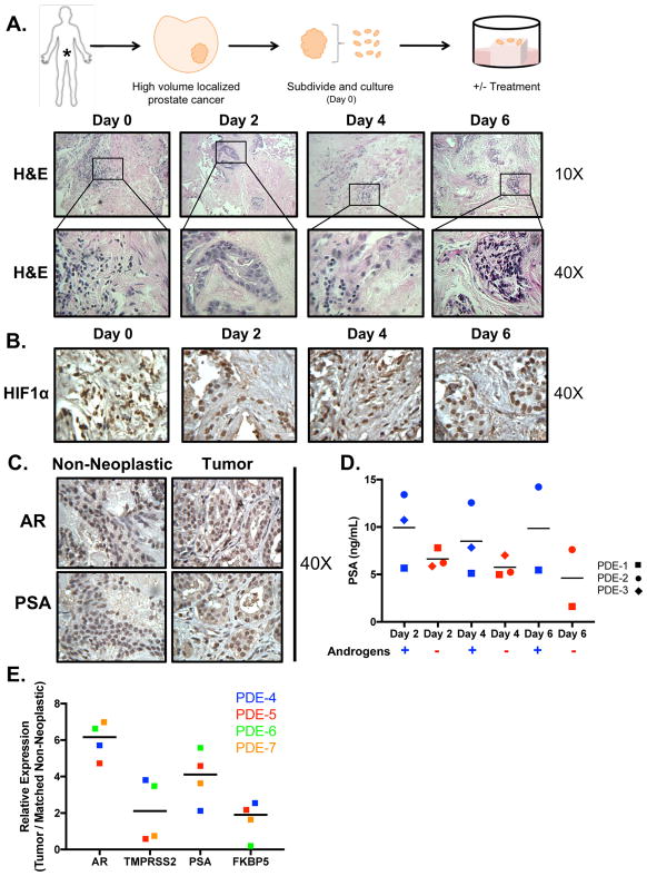
Figure 1. PCa PDE model sustains tumor morphology, viability, and endogenous endocrine signaling.
A. Top: Depiction of the method used to culture patient tumors ex vivo. Drug treatment can be added to the media to investigate the effect on tumor growth. Media (and appropriate treatment) were replaced every other day. Tissue was harvested and fixed in 4% formalin. The formalin-fixed tissue was then embedded into paraffin blocks and cut into sections with a microtome. Bottom: Slides were stained with hematoxylin and eosin (H&E). The representative images shown (10× and 40×) indicate maintenance of gross tumor morphology after culturing ex vivo for up to 6 days. B. PDE were stained for HIF1α showing that the tumors received sufficient oxygen supply to maintain viability. C. AR and PSA immunostaining of (patient #5) tissue demonstrated sustained endogenous and endocrine signaling in the explants after 6 days of ex vivo culture. All IHC images are shown as 40× magnification D. PSA secreted into media of PDE was analyzed at Days 2, 4, and 6 via ELISA in hormone proficient media (‘Androgen Proficient’ – square shape) and hormone-deficient media (‘Androgen Deprived’ – diamond shape). E. Expression of AR and AR target genes (TMPRSS2, PSA, and FKBP5) in explants from four different PDE shown as relative expression normalized to 18S. Tumor tissue was matched to their respective non-neoplastic tissue control.

