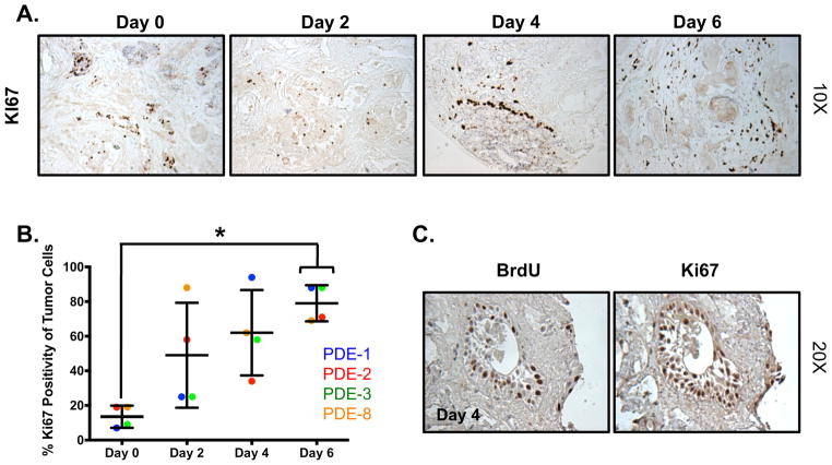
Figure 2. Tumor cells in PDE exhibit de novo proliferative capacity.
A. Tissue was cultured in complete media and harvested every 48 hours for up to 6 days. Ki67 staining was performed to determine the amount of proliferation in the explants. Images are shown at 10X magnification. B. Quantification of Ki67 immunostaining showed a time-dependent increase in proliferation. PDE 3 is shown in panel A. *p<0.05. C. BrdU uptake is similar to Ki67 staining. Images are shown at 20X magnification, n=4.

