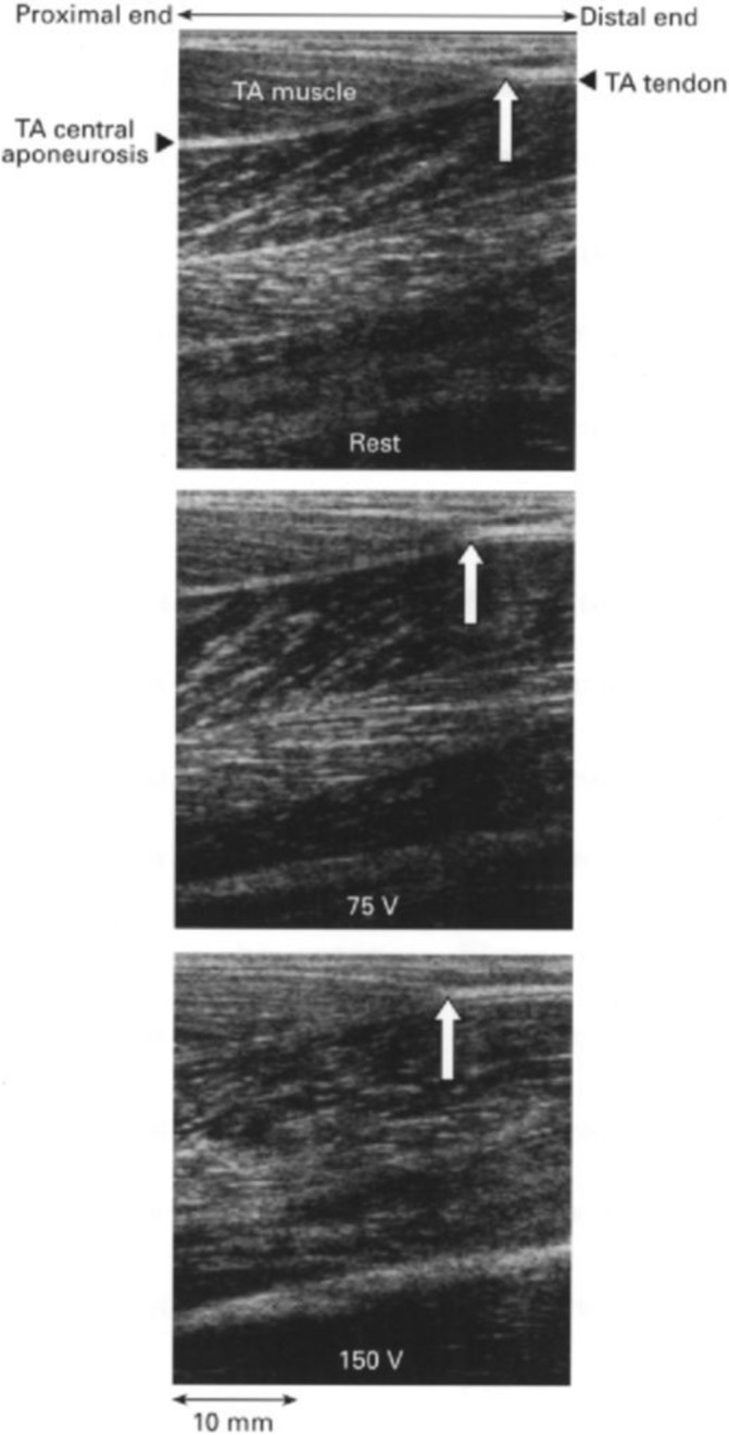
Figure 5.

Tendon displacement measured by B-mode ultrasound. Sonographic images of the human tibialis anterior (TA) muscle at rest (top) and in response to electrical stimulation at 75 V (middle) and 150 V (bottom). The white arrow indicates the TA tendon origin. Notice the proximal shift of the TA tendon origin on electrical stimulation.88
