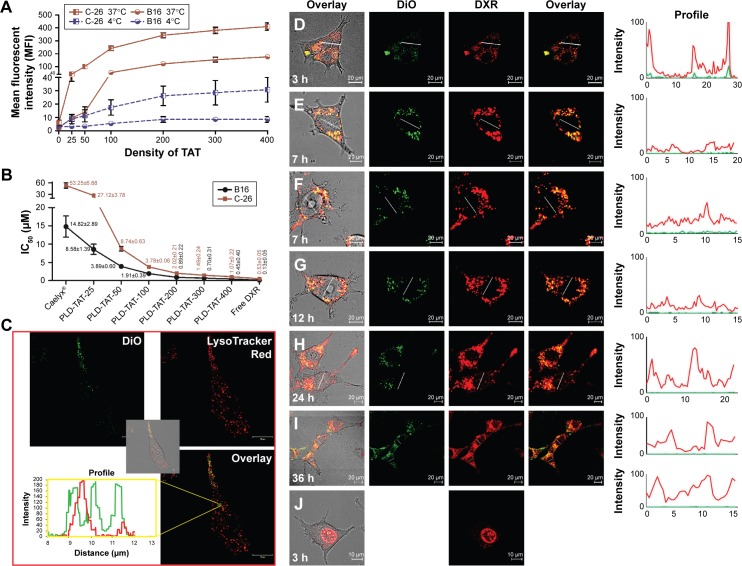
Figure 2.
In vitro analysis of cellular association and cytotoxicity of TAT-modified liposomes.
Notes: Panel A represents the association of different FPLs with C26 and B16 cells at 37°C and 4°C. Cells (105 cells/well) were exposed to different FPLs (100 nmol phospholipid/500 µL) for 3 hours at either 37°C or 4°C, detached, and association of liposomes with cells was analyzed by flow cytometry. Data depict mean fluorescent intensity ± SD (n=4). Panel B represents the IC50 values of different PLDs against C26 and B16 cells after 3 hours of exposure at 37°C followed by 72 hours of proliferation. Data are expressed as mean ± SD (n=4). Panel C illustrates internalization of FPL-200 into C26 cells pre-incubated with LysoTracker®Red and the profile view of DiO and LysoTracker®Red along a line passed through red and green spots inside the cell after 3 hours of incubation at 37°C. Images D–J illustrate the intracellular fate of DXR delivered to C26 cells at 37°C by PLD-TAT200 (D–I) or free DXR (J) after 3 hours of exposure to preparations and wash. Images were captured at different times after cell exposure to preparations.
Abbreviations: TAT, transactivator of transcription; PLD, PEGylated liposomal doxorubicin; FPL, fluorescent-labeled PEGylated liposome; DiO, 3,3′-dioctadecyloxacarbocyanine perchlorate; DXR, doxorubicin.

