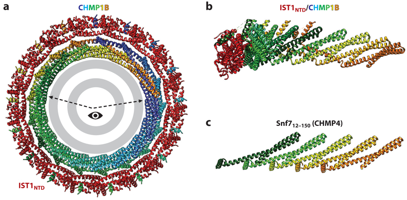
Figure 3. ESCRT-III filament structures.
(a) End-on view of a turn of the N-terminal ESCRT-III domain of an IST1NTD/CHMP1B filament surrounding a stylized lipid bilayer. IST1NTD subunits are shown in red, CHMP1B subunits are shown in rainbow colors, and the lipid bilayer is shown in gray. The structure is from McCullough et al. (2015). (b) Side view of a segment of the IST1NTD/CHMP1B filament (viewed from the membrane and corresponding to the wedge highlighted in panel a). Seven interacting CHMP1B subunits are shown, with just a single associated ISTNTD subunit shown for clarity. (c) Equivalent view showing a linear strand of Snf712–150 and emphasizing the equivalent packing of N-terminal helical hairpins in the two ESCRT-III strands. The structure is from Tang et al. (2015).

