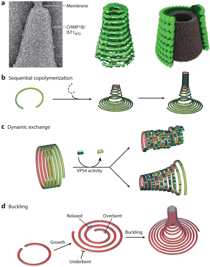
Figure 6. ESCRT-III filament topologies and membrane remodeling.
(a) Cone formation by double-stranded filaments formed by the N-terminal ESCRT-III domain of IST1 (IST1NTD) and CHMP1B. (Left to right) Double-stranded IST1NTD/CHMP1B filaments wrapping about a conical membrane; reconstructed cone comprising double-stranded IST1NTD/CHMP1B filaments; schematic depiction of the structures shown in the left panel, with the IST1NTD strand shown in light green, the CELMP1B strand shown in dark green, and the internal lipid bilayer shown in gray, (b–d) Illustrations of how membrane deformation, constriction, and fission could be driven by heteromeric and dynamic ESCRT-III filaments that adopt different topologies in response to a variety of conditions, including (b) sequential copolymerization of different ESCRT-III subunits with distinct intrinsic curvatures, (c) dynamic exchange of subunits with high intrinsic curvature into tubes of less curved filaments, and (d) out-of-plane buckling induced by the accumulation of elastic stress arising from growth of a spiraling filament beyond its preferred radius of curvature. For simplicity, panels b–d show single long filaments, but analogous principles could also apply to arrays of shorter, close-packed filaments. Illustrations in panel a were adapted from McCullough et al. (2015) and reproduced with permission from AAAS. Illustrations in panels b–d were adapted from Chiaruttini & Roux (2017).

