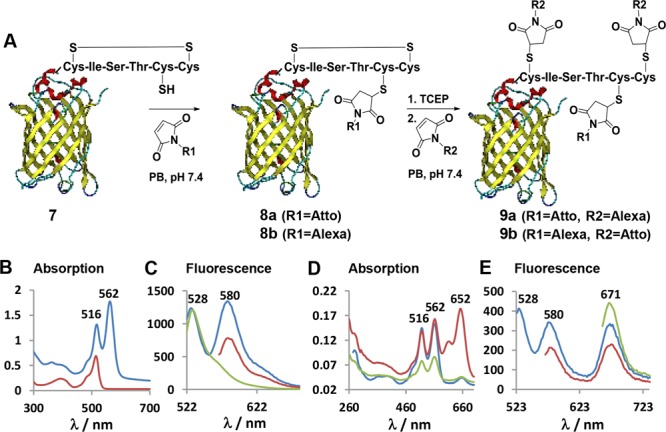
Figure 5.

(A) Site-specific labeling of Dis-tag-EYFP 7 with two different fluorophores denoted as R1 and R2 in a generic fashion. (B) Absorption spectra of Dis-tag-EYFP 7 (red) and Atto550-EYFP 8a (blue) obtained after the first labeling step. (C) Emission spectra of the Atto550-EYFP 8a excited at 516 nm (blue), 562 nm (red), and Dis-tag-EYFP 7 excited at 514 nm (green). (D) Absorption spectra of the dual labeled Atto550-Alexa647-EYFP 9a (red) after the second labeling step and Atto550-EYFP 8a as control that has been incubated with Alexa647-maleimide without preincubation of TCEP (blue). Atto550-EYFP 8a (green) after first capping the additional free cysteine with N-(2-aminoethyl)maleimide and addition of Alexa647 without preincubation of TCEP (green). (E) Emission spectra of the Atto550-Alexa-EYFP 9a excited at 516 nm (blue), 562 nm (red), and 652 nm (green). Absorption and emission spectra of 8b and 9b are given in the Supporting Information Figure S11.
