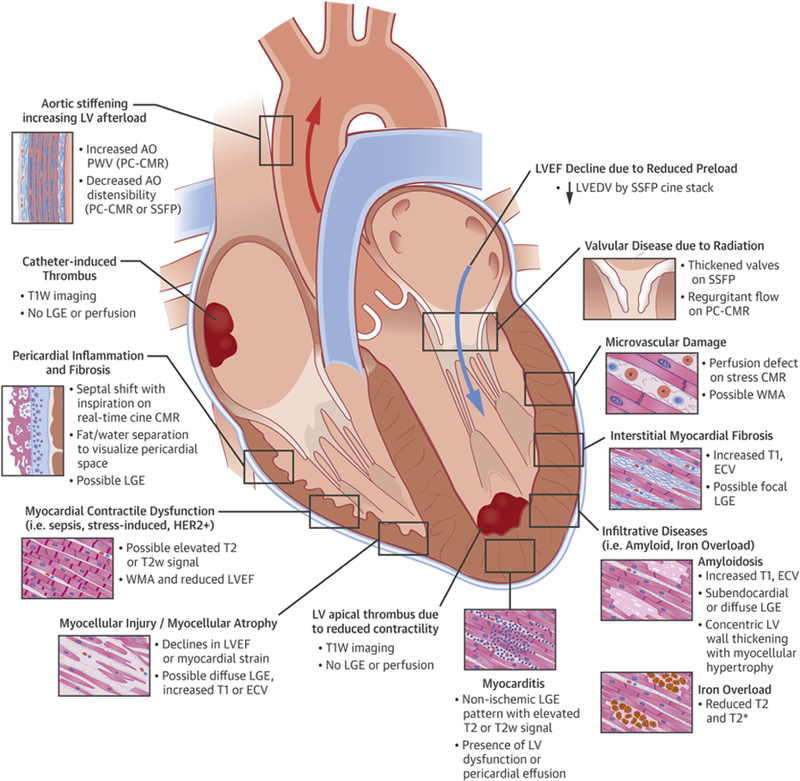
CENTRAL ILLUSTRATION.
Adverse Cardiovascular Effects Related to Cancer Treatment and Key Cardiovascular Magnetic Resonance Features

This figure illustrates cardiovascular complications that may be found in patients with cancer or survivors related to their cancer treatment. Cardiovascular magnetic resonance (CMR) imaging may be useful to not only identify these disease processes but also comprehensively assess their impact on cardiovascular function. AO = aorta; ECV = extracellular volume; HER2 = human epidermal growth factor receptor 2; LA = left atrium; LGE = late gadolinium enhancement; LV = left ventricle; LVEDV = left ventricular end-diastolic volume; LVEF = left ventricular ejection fraction; PA = pulmonary artery; PC = phase-contrast; PWV = pulse-wave velocity; RA = right atrium; RV = right ventricle; SSFP = steady-state free precession; T1W = T1-weighted; T2w = T2-weighted; WMA = wall motion abnormality.
