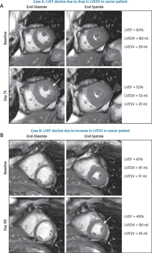
FIGURE 3.
Cardiovascular Magnetic Resonance Case Examples of Left Ventricular Ejection Fraction Due to Either a Decline in Left Ventricular End-Diastolic Volume or an Increase in Left Ventricular End-Systolic Volume After Chemotherapy

Cardiovascular magnetic resonance images demonstrating mid-short-axis white-blood slice acquired prior to treatment (top) and 3 months post-treatment (bottom), with end-diastolic frames shown on the left and end-systolic frames shown on the right. (A) A case of a precipitous decline in left ventricular ejection fraction (LVEF) after 3 months of treatment due to a decline in left ventricular end-diastolic volume (LVEDV) of unknown origin in a 57-year-old white woman with a history of diabetes and hypertension who was treated with 2 cycles of sunitinib for metastatic renal cell carcinoma. As shown in this case example, the baseline LVEF was 63% (Online Video 1) and declined to 52% (Online Video 2) 75 days thereafter because of a change in LVEDV. (B) A case of anthracycline-mediated LV systolic dysfunction with a precipitous decline in LVEF 3 months after initiation of treatment. In this situation, LV end-systolic volume (LVESV) increased in a 59-year-old white woman with a history of smoking who was treated with anthracycline-containing chemotherapy for breast cancer. As shown in this case example, the baseline LVEF was 61% (Online Video 3) and declined to 49% (Online Video 4) 99 days thereafter because of a change in LVESV.
