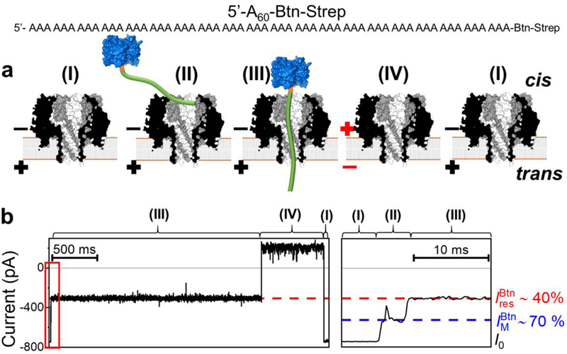
Figure 4.

Capture and immobilization of A60-Btn-Strep in γ-HL. (a) Schematic showing: (I) open channel of γ-HL; (II) electrophoretically captured ssDNA exploring the vestibule; (III) ssDNA threading through the β-barrel; (IV) release of ssDNA by reversing the potential polarity. The ssDNA [poly(dA)60] (green) is attached to streptavidin (blue) using a biotin linker (orange). (b) Left: sample I-t trace labeled with proposed steps, (I)-(IV). Right: expanded view of the mid-level blocking current indicated in the red box. Recordings were carried out at −120 mV, in 3 M KCl, 20 mM HOAc/KOAc, pH 5.0, at 20 °C. The I-t traces were post-filtered at 2 kHz for presentation. Open-channel current (I0), mid-level blockade (), and deep current blockade correspond to (I), (II), and (III), respectively.
