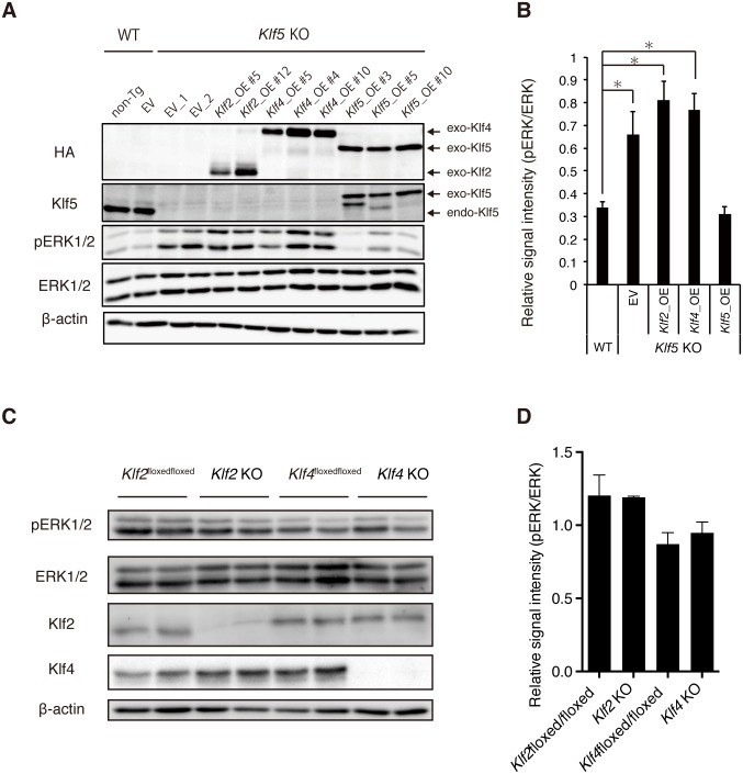
Fig 2. Rescue of elevated pERK levels in Klf5-KO ESCs by Klf5, but not Klf2 or Klf4.
(A) Level of pERK in Klf5-KO ESCs overexpressing Klf2, Klf4, or Klf5. Western blot analysis of WT non-transgenic (non-Tg), empty vector (EV) control, Klf5-KO EV control, Klf2-overexpressing (Klf2_OE), Klf4_OE, and Klf5_OE ESCs were performed with antibodies against HA, Klf5, pERK, ERK, and β-actin. Exo; exogenous, endo; endogenous. (B) Quantified signal intensity for pERK relative to ERK. (C) Level of pERK in Klf2-or Klf4-KO ESCs. Western blot analysis of WT, Klf2-KO ESCs, and Klf4-KO ESCs. (D) Quantified signal intensity of pERK relative to ERK. Asterisk indicates statistical significance: *P < 0.01; Mann-Whitney U test.

