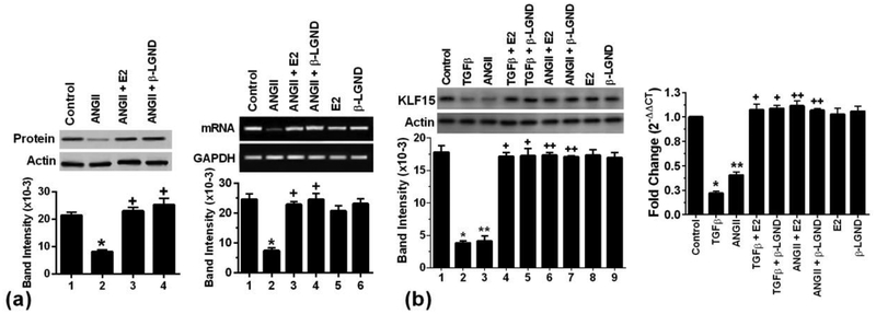
Fig 1. ERβ opposes ANGII suppression of KLF15 expression.
(a) AngII (100nM) inhibits and E2 (10nM) or β-LGND (10nM) prevents the inhibition of KLF15 protein (left), and mRNA (right) expression in cultured cardiomyocytes. Actin and GAPDH, respectively, are loading controls. Bar graph is mean± SD from 3 exps. p<0.05 vs control, +p<0.05 vs AngII alone from analysis by ANOVA + Schefe’s test. (b) Equimolar TGFβ (10ng/ml) or AngII represses KLF15 protein (left) and mRNA (right) that was significantly prevented by E2 or β-LGND. *p<0.05 or **p<0.05 vs control, +p<0.05 or ++p<0.05 vs AngII or TGFβ alone, n=3 exps.

