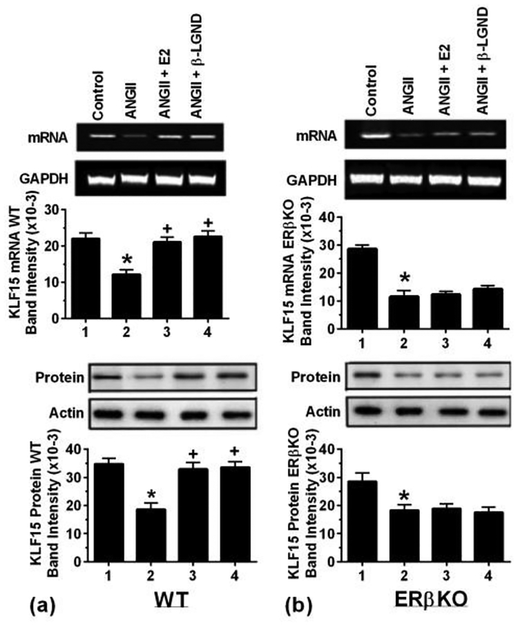
Fig 6-. In-vivo studies of AnglI-induced cardiac hypertrophy.
Ovexed, female mice were infused with saline or AnglI± β-LGND or E2 (pellet) for 4 weeks (4 mice per condition), and the hearts were processed for KLF15 mRNA and protein. Results are from pooled (a) WT mice and (b) ERβKO mice. Actin and GAPDH, respectively, are loading controls. Data are from 4 mice, pooled ventricles per condition for the Figs, each in WT and ERβKO mice. Bar graph data are analyzed by ANOVA and Scheffe’s test. *p < 0.05 vs. control, +p < 0.05 for AngII versus same + E2 or β-LGND.

