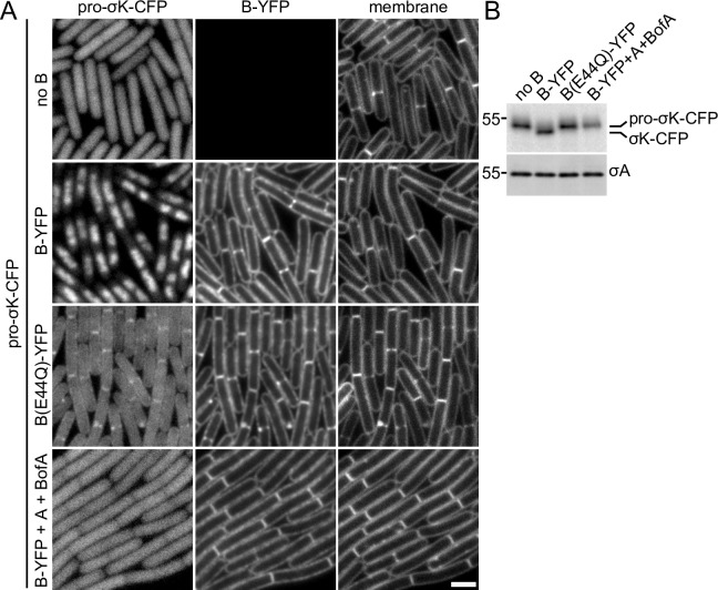
Fig 4. Pro-σK-CFP localizes to the membranes of vegetatively growing cells when B(E44Q) is co-expressed.
(A) Representative images of the indicated strains after 1.5 hours of induction. In the absence of the B protease, pro-σK-CFP localizes in the cytoplasm. In the presence of wild-type B-YFP, pro-σK-CFP gets processed and localizes to the nucleoid. Pro-σK-CFP co-localizes with B(E44Q)-YFP in the septal and cytoplasmic membranes when co-expressed during vegetative growth. Pro-σK-CFP remains cytoplasmic when wild-type B-YFP is co-expressed with its inhibitors A and BofA. The cytoplasmic Pro-σK-CFP signal is weaker and more pixelated in the strain expressing B(E44Q)-YFP compared to those lacking B or co-expressing A and BofA (see text). All images were scaled identically. Larger fields of cells over the induction time course can be found in S6 Fig. Scale bar indicates 2 μm. (B) Immunoblot of the same strains in (A) using anti-σK antibodies showing pro-σK-CFP processing in cells expressing B-YFP but not in cells expressing the E44Q mutant, the inhibitors A and BofA, or in a strain that does not express B during vegetative growth. σA was used to control for loading. Molecular weight markers (in kDa) are indicated to the left. An anti-GFP immunoblot to assess the relative amount of liberated (free) CFP in the four strains can be found in S2C Fig.

