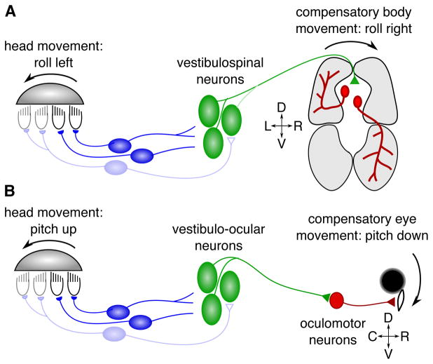
Figure 2. Schematic of vestibular circuits subserving posture and gaze stabilization.
A) At the onset of movement, the utricular otolith slides relative to the hair cells underneath, depolarizing some (black) and hyperpolarizing others (gray), depending on their ciliary orientation. Vestibular afferents (blue) relay hair cell signals to vestibulospinal neurons in the hindbrain (green). This vestibular drive sets up asymmetric activation of trunk musculature through as-yet unclear connectivity that is thought to rely both on direct synapses with motor neurons as well as indirectly via spinal premotor populations (Kasumacic et al., 2015; Murray et al., 2018). In this example, as the fish rolls to the left, stronger motor drive to ventral muscle on the right and dorsal muscle on the left (gray shaded regions) produces a self-righting torque (Bagnall and McLean, 2014). B) Equivalent schematic for vestibular-driven gaze stabilization. Here the brainstem vestibular nuclei are known to make direct connections to oculomotor neurons. Recent work has revealed that in pitch-related circuits, vestibulo-ocular neurons driving eye rotation downwards (active pathway, dark green) outnumber those driving upwards eye movements (inhibited pathway, pale green) by 6:1 (Schoppik et al., 2017).

