
Figure 2. IL-33 was not elevated in NPs.
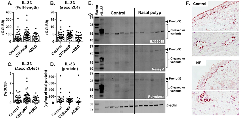
Expression of mRNAs for full-length IL-33 (A), IL-33∆exon3,4 (B) and IL-33∆exon3,4s5 (C) was determined by RNase H-dependent quantitative RT-PCR in control ethmoid sinus tissue (Control; n = 34) and NPs from CRSwNP (n = 46) and AERD (n = 23). IL-33 protein in tissue extracts was determined by Luminex (D, control (n = 26), CRSwNP NP (n = 37) and AERD NP (n = 13)) and Western blot (E). IL-33 protein concentration was normalized to the concentration of total protein (D). The results are representative of 2 separate experiments with separate donors (control (n = 12) and NP (n = 11)) (E). Representative immunostaining for IL-33 is shown in a control ethmoid tissue and an NP tissue (F). Magnification; ×400 (upper) and ×1200 (lower). Results are shown as median with interquartile range.