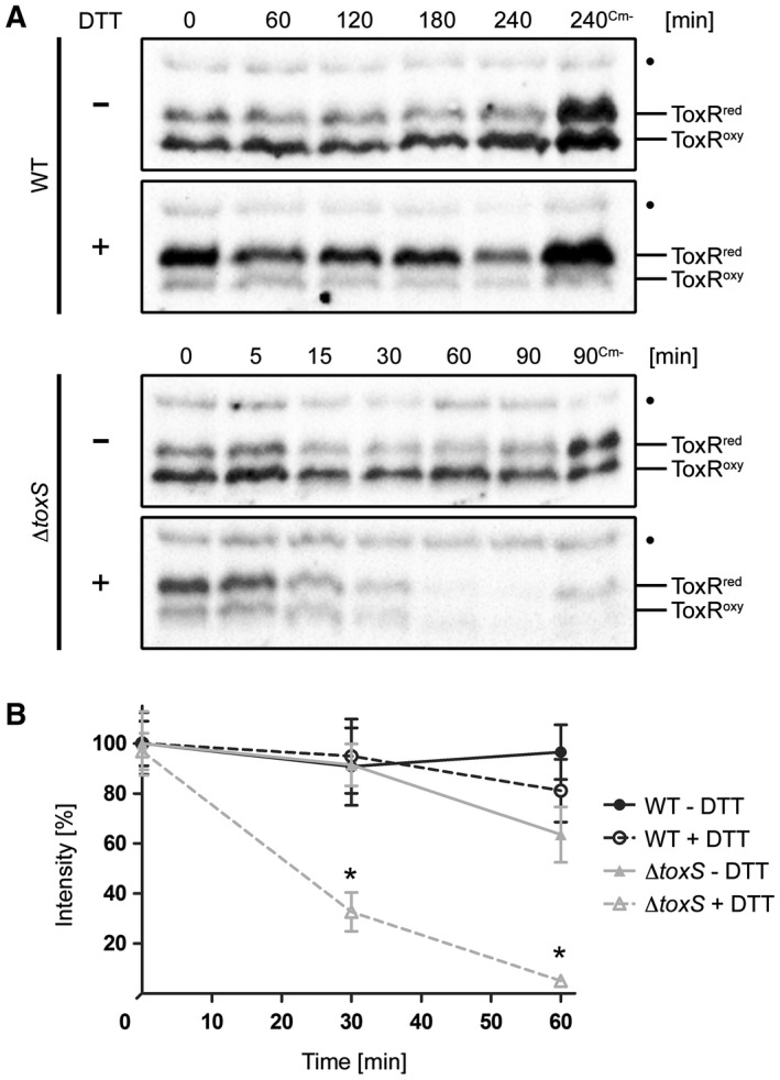
Figure 2.

Effects of DTT treatment on the redox state and protein stability of ToxR in V. cholerae WT and toxS mutants. ToxR temporal stability levels were measured by the immunoblot analysis in WT and toxS mutant strains grown in M9 maltose with or without DTT (+/–). Protein biosynthesis was inhibited by the addition of Cm. Samples without chloramphenicol (Cm‐) served as negative controls. A. Immunoblots of WT and toxS WCL were performed under standard nonreducing Laemmli buffer conditions utilizing α‐ToxR antiserum. The migration patterns of ToxRred/oxy are indicated. (•): Represents a nonspecific cross‐reacting background band. B. Graphs show band intensities (%) of WCL samples treated under reducing Laemmli buffer conditions defined by densitometry of similar blots (see one set of representative immunoblots Fig. S4). For each time point, the sample number was n ≥ 6 and the mean values with standard deviation are shown. Two‐way ANOVA with Bonferroni post hoc analysis indicates significant differences between toxS strains without (grey filled triangle, solid line) and toxS cells with DTT (grey open triangle, dotted line) with P < 0.001 at time points 30 and 60 min. No significant differences were seen between WT without DTT (black filled circle, solid line) and WT incubated with DTT (black open circle, dotted line).
