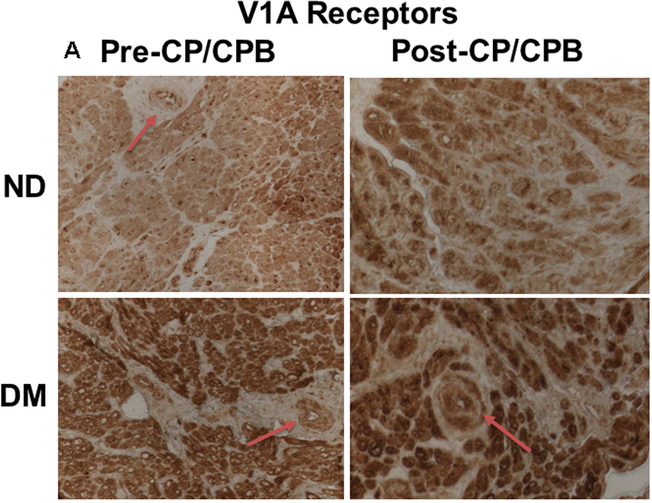
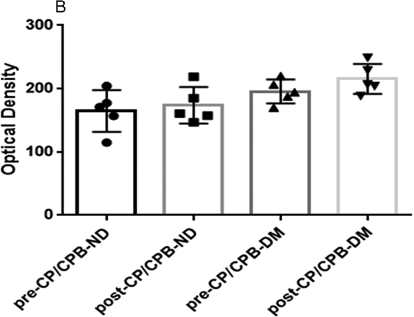
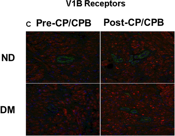
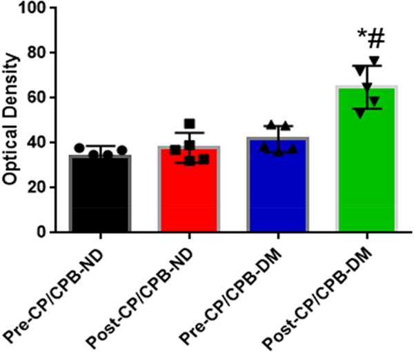
Figure 4.
A, Immunohistochemical image of V1A receptors in paraffin-embedded human atrial tissue slides from non-diabetic (ND) and diabetes (DM) patients. B, Densitometric analysis of signal intensities, C, Immunofluorescence staining of V1B receptors in the embedded human atrial tissue slides from the ND and DM patients before and after CP/CPB. D, Densitometric analysis of signal intensities. *P<0.05 vs. pre-CP/CP-DM, #P < 0.05 vs. post-CP/CPB-ND; n = 5/group, mean ± SD.


