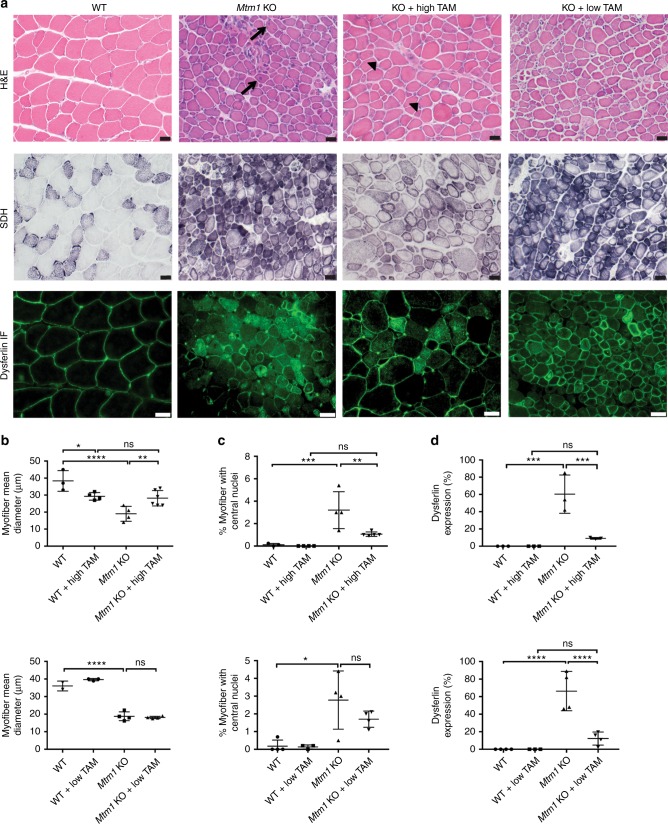
Fig. 2.
Tamoxifen treatment improves Mtm1 knockout mouse muscle structure. a Tibialis anterior muscle stained with hematoxylin and eosin (H&E) and succinate dehydrogenase (SDH), and by anti-dysferlin immunofluorescence (IF). Untreated Mtm1 KO muscle has centrally located nuclei (arrows in H&E), mitochondrial aggregation on SDH staining, and abnormal distribution of dysferlin staining. High-dose TAM treatment in KOs results in: (1) reduction of central nuclei (arrowheads); (2) improvement in myofiber size; (3) resolution of mitochondrial aggregation; and (4) restoration of dysferlin to the sarcolemmal membrane. Low-dose TAM improves dysferlin localization but does not resolve central nucleation and myofiber hypotrophy. All drug treatments started at 21 days, and histopathology performed at 36 days of age. b High-dose TAM treatment increases myofiber size of Mtm1 KOs. TAM treated WT (29 ± 3 μm, n = 4) vs. untreated Mtm1 KO (19 ± 3 μm, n = 4, ****p < 0.0001 vs. WT), vs. TAM treated KO (28 ± 3 μm, n = 6, **p = 0.0065 vs. untreated KO, not significant vs. WT + TAM). c High-dose TAM treatment reduces % central nuclei in Mtm1 KOs (per 100 fibers): TAM treated WT (0%, n = 4) vs. untreated Mtm1 KOs (3 ± 0.8%, n = 4, ***p = 0.0004 vs. WT), vs. TAM treated KOs (1.1 ± 0.09%, n = 5, **p = 0.0024 vs. KOs and n.s. vs. WT + TAM). d High and low-dose TAM treatment restores dysferlin localization to cell membrane. Untreated Mtm1 KO = 60 ± 13% cytoplasmic (n = 3, ***p = 0.0002 vs. WT), KO + high TAM = 9 ± 1% cytoplasmic (n = 3, ***p = 0.0005 vs. KO, ns compared to WT + TAM). For low-dose TAM, Mtm1 KO = 66 ± 11% cytoplasmic, (n = 3, ****p = < 0.0001 vs. WT) vs. KO + low TAM = 12 ± 4% cytoplasmic (n = 4, ****p < 0.0001 vs. KO, ns compared to WT + TAM). All data points for b–d presented in Supplementary Table 2. Statistical analyses were conducted by two-way ANOVA, followed by Tukey’s multiple comparisons post-test or Fisher’s Least Common Differences post-test. For direct two sample comparison, unpaired, parametric two-tailed Student's t-test was performed. Scale bars = 20 µm

