Fig. 1.
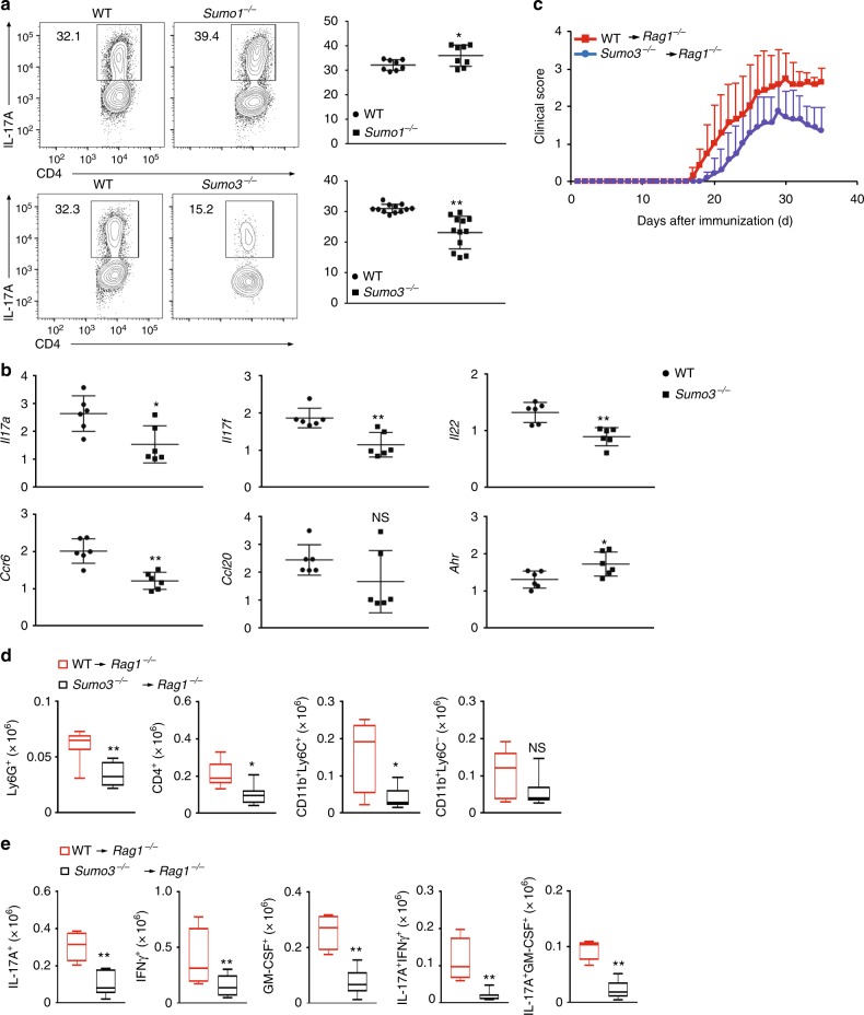
SUMO3, but not SUMO1, stimulates TH17 differentiation. a Representative flow cytometric analysis of intracellular IL-17A expression (boxed) in naive CD4+ T cells from WT, Sumo1−/− (top), and Sumo3−/− (bottom) mice, cultured in vitro for 3 d under TH17 priming conditions. Numbers adjacent to the outlined area indicate the percentage of the cells in gated area (throughout). b qPCR analysis of Il17a, Il17f, Il22, Ccr6, Ccl20, and Ahr mRNA in WT and Sumo3−/− TH17 cells assessed in (a). Expression is presented relative to that of the control gene Actb. c Mean clinical EAE scores of Rag1−/− mice adoptively transferred with WT or Sumo3−/− CD4+ T cells (key; n = 5 per genotype) from days 0 to 35 after immunization with the EAE-inducing epitope MOG35-55. d Quantification of CNS-infiltrating cells from Rag1−/− mice reconstituted with CD4+ T cells from WT or Sumo3−/−mice (same as in c) expressing characteristic mononuclear cell surface markers, assessed using flow cytometry at the peak of disease. e Flow cytometric analysis of CNS-infiltrating cells from Rag1−/− mice reconsituted with WT or Sumo3−/− CD4+ T cells (same as in c) positive for intracellular cytokines IL-17A+, IFNγ+, GM-CSF+, IL-17A+ IFNγ+, and IL-17A+ GM-CSF+. NS, not significant (P > 0.05); *P < 0.05 (t-test); **P < 0.01 (t-test). Data are from three experiments (a, right; and b–e; presented as median [central line], maximum and minimum [box ends], and outliers [extended lines]) or are from one representative of three independent experiments (a, left)

