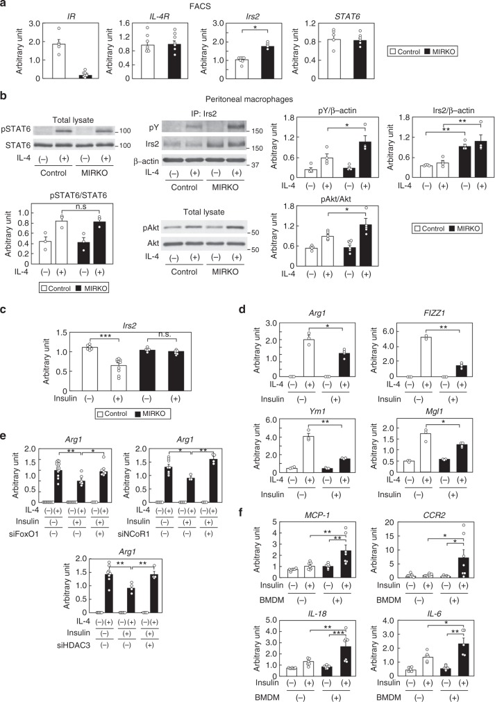
Fig. 5.
The Irs2 mRNA levels and number of M2a-subtype MΦs were elevated in HF diet-fed MIRKO mice. a Expression levels of IR, IL-4R, Irs2 and STAT6 in the siglecF-CD11b+F4/80+ cells of the SVF of the adipose tissue from the control and MIRKO mice (n = 4–6). b IL-4-induced STAT6, Irs2 and Akt phosphorylation in peritoneal MΦs of HF diet-fed MIRKO mice (n = 3–6). c Irs2 expression levels in the BMDM of the control and MIRKO mice after 100 nM insulin stimulation for 3 h (n = 3–4). d IL-4-induced M2a-subtype marker genes in the BMDM after 100 nM insulin pretreatment for 8 h (n = 3–4). e IL-4-induced Arg1 expression levels in the BMDM pretreated with 100 nM insulin for 8 h after siFoxO1, siNCoR1, or siHDAC3 treatment (n = 4–16). f MCP-1, CCR2, IL-18 and IL-6 expression levels in 3T3-L1 cells in co-culture with BMDM of the C57BL/6 mice and 3T3-L1 cells after insulin stimulation for 24 h (n = 7). The data are mean ± SEM. followed by one-way ANOVA with a post hoc test or Student’s t test. *P < 0.05; **P < 0.01; ***P < 0.001

