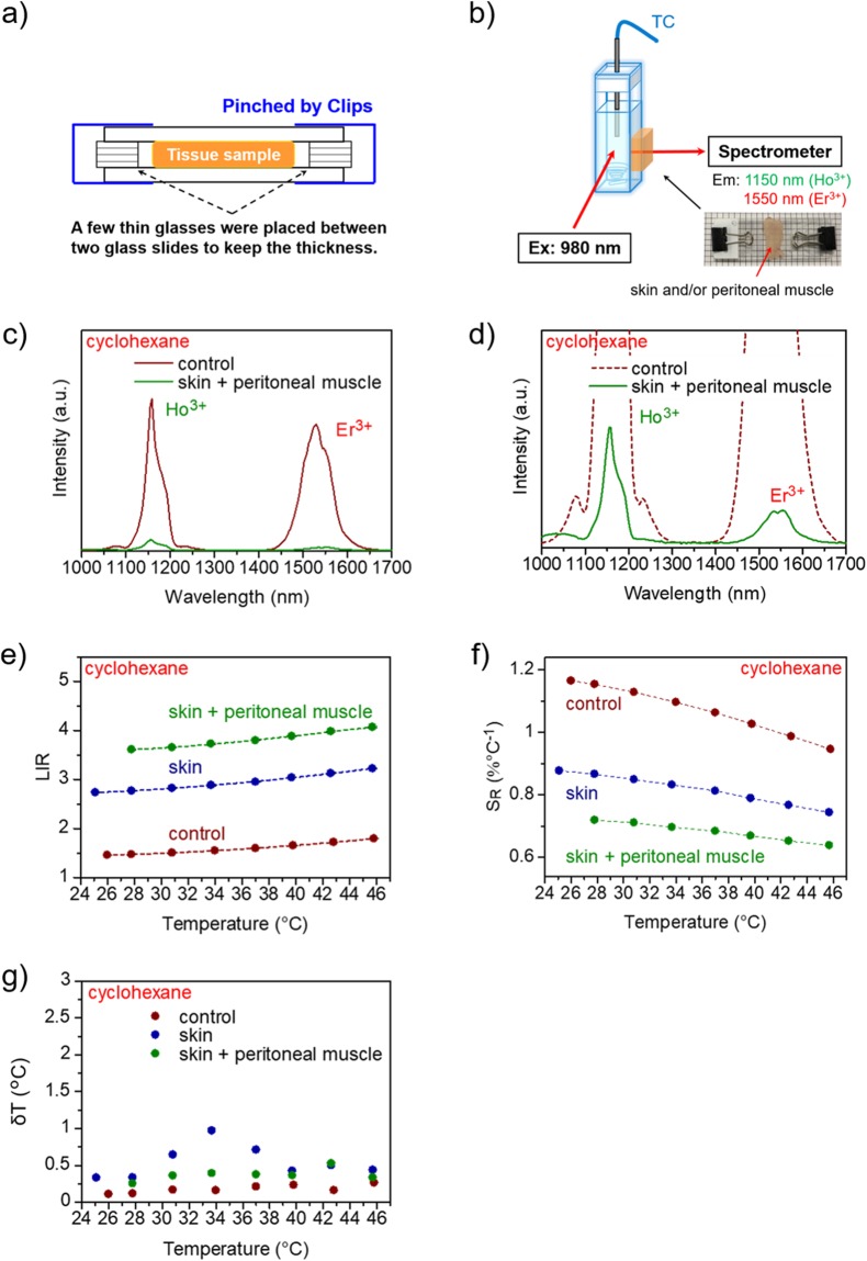
Figure 4.
Temperature-dependent OTN-NIR emission spectra of NaYF4 NPs recorded using biological tissues (skin and/or peritoneal muscle). (a) Schematic representation of the biological tissues used for obtaining the emission spectra. Tissue samples were placed between two glass slides, with the gap maintained at 880 (skin) or 1330 μm (skin + peritoneal muscle). (b) Schematic illustration of recording temperature-dependent emission spectra with biological tissues. The tissue samples were placed between the cuvette and detector of the spectrometer. (c) Comparison of the luminescence intensity of the NaYF4 NPs only (control) vs. that with skin + peritoneal muscle and (d) enlarged view of c. (e) Comparison of the LIR of the NaYF4 NPs (control, skin, and skin + peritoneal muscle). (f) Comparison of the relative thermal sensitivity (SR) and (g) the temperature uncertainty for NaYF4 NPs using skin and peritoneal muscle. The NPs were dispersed in cyclohexane at a concentration of 30 mg/mL. The laser power was 4.22 W.

